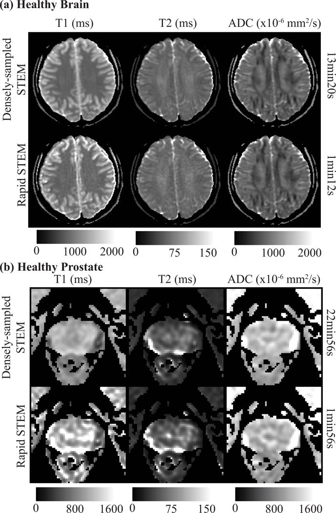
Figure 5.

An example of T1, T2 and ADC maps re-estimated retrospectively with the optimized rapid acquisition protocol (rapid STEM) are shown for healthy brain (a) and healthy prostate (b). The overall measurements are accurate even though the maps are noisier. The T1 map of healthy prostate shows some artifacts, likely due to motion without antiperistatic agents.
