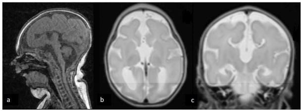
Figure 1abc.
MRI findings at term equivalent age. T1-weighted sagittal view (a) demonstrates cerebellopontine hypoplasia and micrognathia. T2-weighted transverse view (b) demonstrates enlarged occipital horns and agenesis of the corpus callosum with large third ventricle. T2-weighted coronal view (c) demonstrates typical ventricular configuration for agenesis of the corpus callosum with superior extension of an enlarged third ventricle. All three views demonstrate microcephaly and markedly delayed folding pattern for age.

