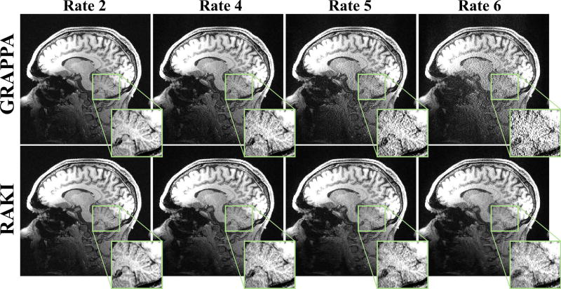
Figure 9.
A central slice of the high-resolution (0.7 mm isotropic) MPRAGE acquisition at 3T, acquired at R = 2 and 5, as well as retrospective R = 4 and 6. At rates 2 and 4, GRAPPA (top) and RAKI (bottom) methods both successfully reconstruct the image with little residual artifacts. At rates 5 and 6, noise amplification becomes visible for GRAPPA reconstruction. At these rates, RAKI has improved noise tolerance and exhibits no blurring artifacts.

