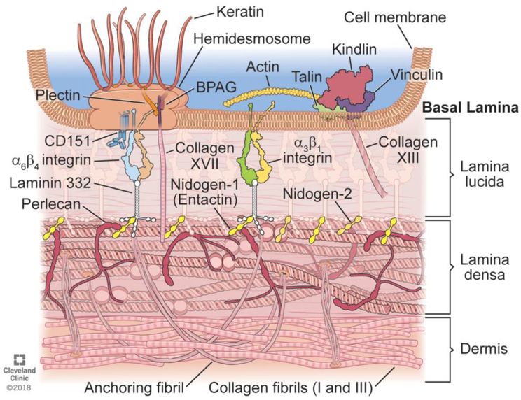
Fig. 1.
Schematic diagram of typical components found in basement membranes, using skin as an example. A basal keratinocyte adheres to the underlying basement membrane and dermis via the focal adhesions that transmit mechanical force and regulatory signals that consist of numerous interacting components such as the hemidesmosome with bullous pemphigoid antigen (BPAG), integrin a6b4, laminin 332, perlecan, anchoring fibrils, and dozens of other components that vary depending on the organ and the status (homeostasis, post-injury, etc.) of the tissues. Many of these components extend into, and are part of, the lamina lucida of the basement membrane. The underling lamina densa of the basement membrane is composed of collagen type IV, nidogens, perlecan, laminin 332, that directly interact with each other, and other components. Lamina lucida is not as wide naturally as it is drawn here for clarity reasons. Illustration by David Schumick, BS, CMI. Reprinted with the permission of the Cleveland Clinic Center for Medical Art & Photography © 2018. All Rights Reserved.

