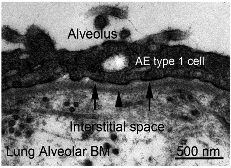
Fig. 6.
Transmission electron micrograph of alveolar basement membrane (BM) of rabbit lung at 46,000X. Shown is the “thicker side” of the alveolus where the alveolar BM and capillary BM are separated by an interstitial space containing collagen fibrils and other extracellular matrix materials. The alveolar epithelial (AE) type 1 cell rests on the BM with lamina lucida (arrows) and lamina densa (arrowhead). On the “thinner side” of the alveolus (not shown) the alveolar BM and capillary BM fuse, at least focally, to form a single BM separating alveolar epithelial type 1 cells and capillary endothelial cells—a BM morphological variation that is thought to facilitate gas exchange between the alveolar space and the alveolar capillaries (Vaccaro and Brody, 1981).

