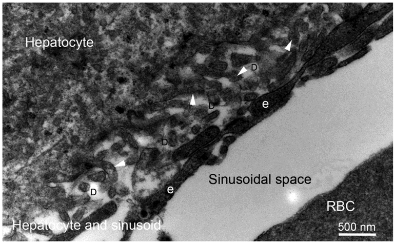
Fig. 7.
Transmission electron micrograph of rabbit liver hepatocyte, sinusoids and spaces of Disse at 30,000X magnification. Hepatocytes are organized into plates separated by the space of Disse (D) from vascular channels termed sinusoids. Hepatocyte processes (arrowheads) extend into the space of Disse. Sinusoids have a discontinuous, fenestrated endothelial cell lining. Of note, there is no basement membrane between either hepatocytes or endothelial cells and the space of Disse—allowing direct cellular contact that is thought to facilitate hepatocyte functions such as detoxification, modification, and excretion of exogenous and endogenous substances.

