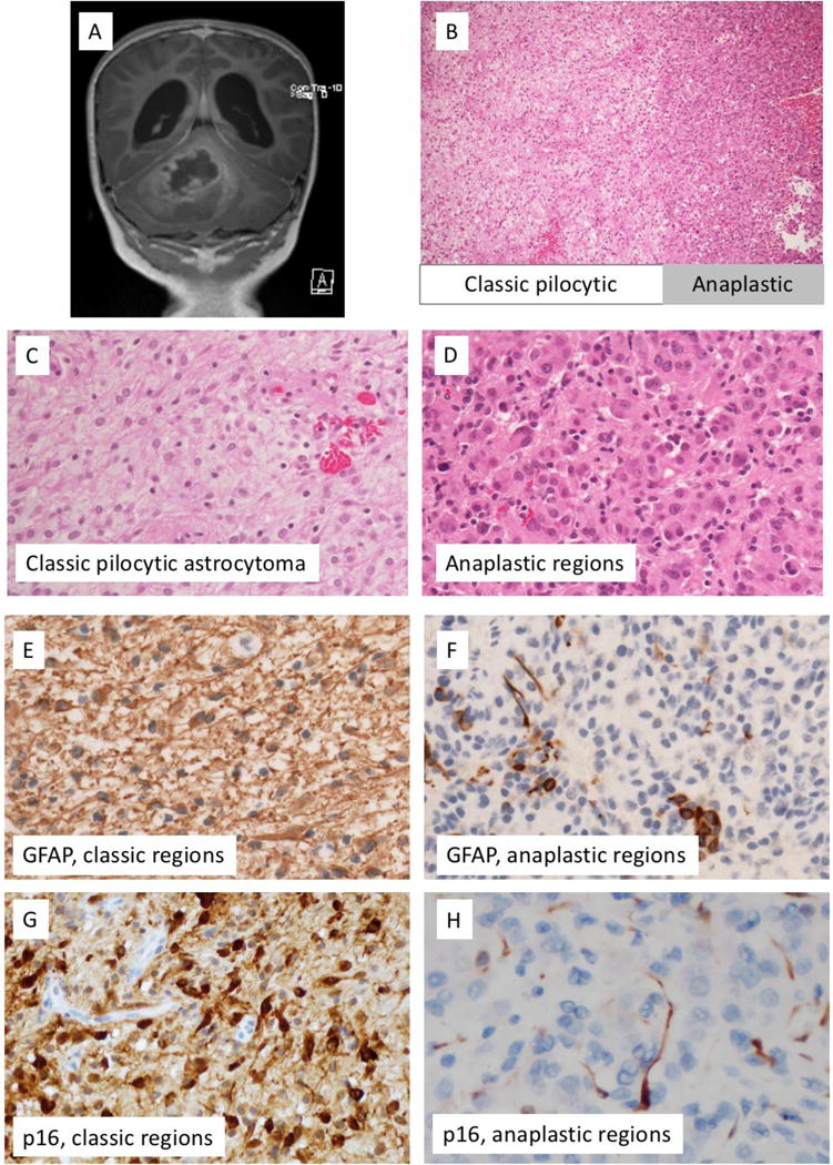
Figure 1.

Findings at time of initial resection. A) Pre-resectionT1-postgadolinium magnetic resonance imaging, B) H&E-stained section, interface of classic-appearing and anaplastic-appearing regions. C) Higher resolution H&E-stained section, region with classic features of pilocytic astrocytoma. D) Higher resolution H&E-stained section, anaplastic region. E) GFAP immunohistochemical staining shows strong diffuse positivity in classic pilocytic regions (E), but only scattered positive cells in anaplastic regions (F). On p16 immunohistochemical stains, the regions with classic features of pilocytic astrocytoma demonstrate diffuse positivity (G), while anaplastic regions demonstrate loss of p16 expression (H).
