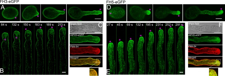
Fig 4. Intracellular localization of AtFH3 and AtFH5 in pollen tubes.
(A) Distribution of AtFH3-eGFP in ungerminated and germinated pollen derived from AtFH3pro:FH3-eGFP;fh3-1 plants. Medial optical sections are presented. The magenta asterisks indicate the localization of AtFH3-eGFP on the PM. Bar = 10 μm. (B) Time-lapse images of AtFH3-eGFP in the growing pollen tube. The magenta asterisks indicate the localiztion of AtFH3-eGFP on the PM and vesicles. Bar = 5 μm. (C) Co-localization of AtFH3-GFP with FM4-64-stained plasma membrane and endocytic vesicles. Bar = 10 μm. (D) Distribution of AtFH5-eGFP in ungerminated and germinated pollen derived from AtFH5pro:FH5-eGFP;fh5-2 plants. Medial optical sections are presented. The magenta asterisks indicate the localization of AtFH5-eGFP on the PM. Bar = 10 μm. (E) Time-lapse images of AtFH5-eGFP in the growing pollen tube. The magenta asterisks indicate the localization of AtFH5-eGFP on the PM and vesicles. Bar = 5 μm. (F) Co-localization of AtFH5-GFP with FM4-64-stained plasma membrane and endocytic vesicles. Bar = 10 μm.

