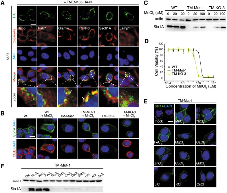
Fig 5. Adding manganese into medium restores binding of Stx1 to TMEM165-deficient cells.
(A) HA-tagged TMEM165 colocalizes with Giantin and TGN46 in 5637 cells. (B) Pretreatment of TM-Mut-1 and TM-KO-3 cells with MnCl2 (100 μM, overnight) restored binding of Stx1 and CtxB to cell surfaces. (C) Binding of Stx1 to TM-Mut-1 and TM-KO-3 cells pretreated with MnCl2 (20 and 100 μM, overnight) was examined by immunoblot analysis, using a polyclonal Stx1 antibody. Actin served as a loading control. One of two independent experiments is shown. MnCl2 did not affect binding of Stx to WT cells but restored binding of Stx1 to TM-Mut-1 and TM-KO-3 cells. (D) WT, TM-Mut-1, and TM-KO-3 cells were exposed to a series of concentrations of MnCl2 for 3 d. Cell viability was determined using MTT assays. TM-Mut-1 and TM-KO-3 cells show greater sensitivity to the toxicity of MnCl2 compared with WT cells (Student’s t test, p = 0.012). Error bars indicate mean ± SD, N = 3. (E-F) Immunofluorescence analysis (E) and immunoblot analysis (F) were carried out to examine metal specificity. TM-Mut-1 cells pretreated with or without a panel of metal chlorides (100 μM, overnight) were subjected to toxin surface-binding assay. MnCl2, FeCl3, and NiCl2 restored binding of Stx1 to cell surfaces. Scale bar, 5 μm. Arrow, colocalization. Representative images were from one of three independent experiments.

