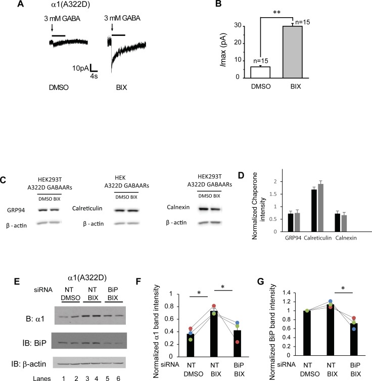
Fig 4. BIX enhances the function of α1(A322D)β2γ2 receptors.
(A) Representative whole-cell patch clamping recording traces in monoclonal HEK293T cells stably expressing α1(A322D)β2γ2 GABAA receptors. Cells were treated with BIX (12 μM, 24h) or DMSO before voltage clamping. GABA (3mM) was applied to induce chloride currents with a holding potential of -60 mV. Quantification of the peak currents (Imax) is shown in (B). The number of patched cells in each group is shown on the top of the bar. pA: picoampere. (C and D) HEK293T cells expressing α1(A322D)β2γ2 receptors were treated with BIX (12μM, 24h). The cell lysates were then subjected to SDS-PAGE and Western blot analysis using corresponding antibodies (C). Quantification of normalized total cellular chaperone protein expression levels is shown in (D) (n = 4, paired t-test). (E) HEK293T cells stably expressing α1(A322D)β2γ2 receptors were treated with non-targeting (NT) or BiP siRNA for 48 hrs. Cells were then treated either with BIX (12 μM) or DMSO vehicle control for another 24 hrs. Cells were then lysed and subjected to SDS-PAGE and western blot analysis. The quantitation results of α1(A322D) and BiP are shown in (F&G) (n = 3, one-way ANOVA). * p < 0.05; ** p < 0.01.

