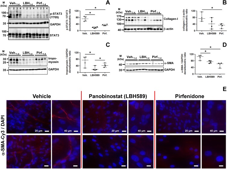
Fig 7. Analysis of STAT3 phosphorylation status and profibrotic protein expression in primary IPF-fibroblasts in response to treatment with panobinostat or pirfenidone.
Primary IPF-fibroblasts (n = 4) were incubated for 24h with vehicle [Veh., 0.25% (v/v) DMSO], panobinostat (LBH589, 85 nM) or pirfenidone (Pirf., 2.7 mM), followed by immunoblot analyses of harvested cells. (A-D) Representative and quantitative immunoblotting for (A) p-STAT3/STAT3, (B) collagen-I, (C) tropomyosin and (D) α-SMA. STAT3, GAPDH or β-actin served as loading control. Data are presented as median ± range of the individual values of different treatments. *p<0.05, by Mann Whitney test. (E) Immunofluorescence for α-SMA-Cy3 (red) on fixed IPF-fibroblasts (n = 3) after vehicle- (left panel), LBH589- (middle panel) or pirfenidone-treatment (right panel). Nuclei were counterstained with DAPI (blue). Representative photographs are shown.

