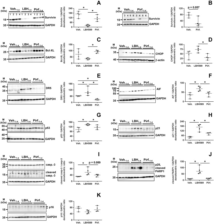
Fig 8. Analysis of pro-apoptotic signaling in primary IPF-fibroblasts in response to treatment with panobinostat or pirfenidone.
Primary IPF-fibroblasts (n = 4) were incubated for 24h with vehicle [Veh., 0.25% (v/v) DMSO], panobinostat (LBH589, 85 nM) or pirfenidone (Pirf., 2.7 mM), followed by immunoblot analyses of harvested cells. (A-K) Representative and quantitative immunoblotting for (A, B) survivin, (C) Bcl-XL, (D) CHOP, (E) DR5, (F) AIF, (G) p53, (H) p21, (I) caspase-3/cleaved caspase-3, (J) cleaved PARP1-p25, and (K) p16. GAPDH or β-actin served as loading control. For survivin-immunoblot (B), additional IPF-fibroblasts (patients 5 and 6) were analyzed. Data are presented as median ± range of the individual values of different treatments. *p<0.05, by Mann Whitney test.

