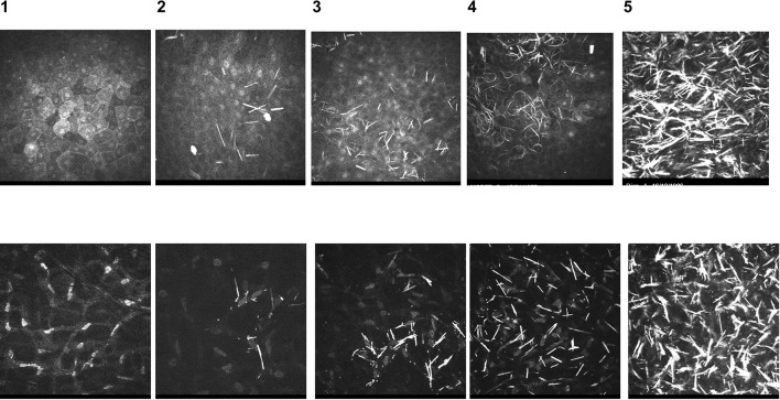
Fig. 2.
In vivo confocal microscopy (IVCM) standardised images used to compare and score images from cystinosis patients. Standardised IVCM images (400 × 400 μm) used to compare and grade images of patient corneal layers, represented in percentages to indicate the number of deposits in the field of each image: 0, no crystal; 1, < 25%; 2, 25–50%; 3, 50–75%; 4, > 75%. Upper panel: superficial epithelium; lower panel: stroma.
Original images provided by H. Liang, Quinze-Vingts National Ophthalmology Hospital, Paris, France

