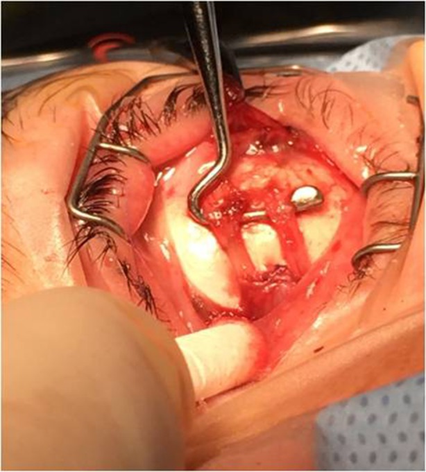
Image 1 Representation of the medial rectus muscle (MR) recession procedure: the central part of the MR tendon and belly are recessed and sutured onto the sclera, leaving intact a 1.5-mm width of the upper and lower tendon insertion pole. (MR shown on the hook in image)
