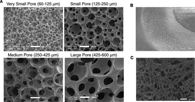
Figure 1.
SEM Micrographs of nanofibrous PLLA scaffolds of four distinct pore size ranges with uniform, spherical, well-interconnected macro-pores (A, scale bar = 200 μm). At higher magnification, the nanofibrous microstructure of the pore walls is apparent (B, scale bar = 10 μm), and an appreciation for their biomimetic diameter is apparent at even higher magnification (C, scale bar = 2 μm).

