Fig. 4.
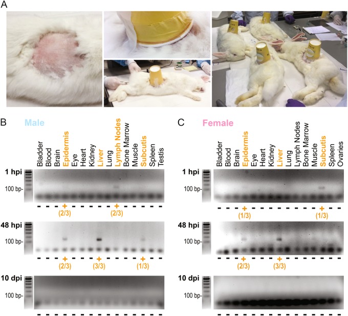
PbVac tissue biodistribution in NZW rabbits. a PbVac administration to rabbits by mosquito bite. Left: a circular area of ~10 cm diameter of the rabbit flank was shaved prior to exposure to mosquitoes. Middle: a container with 100 mosquitoes was placed on the shaved flank of the sedated animal and taped to the skin. Right: the mosquitoes were allowed to feed on the sedated animals for 15 min. b, c Gel electrophoresis analysis of products of qRT-PCR amplification of a 134 bp fragment of the PbVac 18S ribosomal gene in various organs of male (b) and female (c) rabbits at different time points after parasite administration. Orange highlights correspond to organs where PbVac was detected. The numbers correspond to the number of animals where the parasite was detected in a particular organ/the number of animals for which that organ was analyzed. Three biological replicates, each including one male and one female NZW rabbit, were employed per time point

