Figure 5.
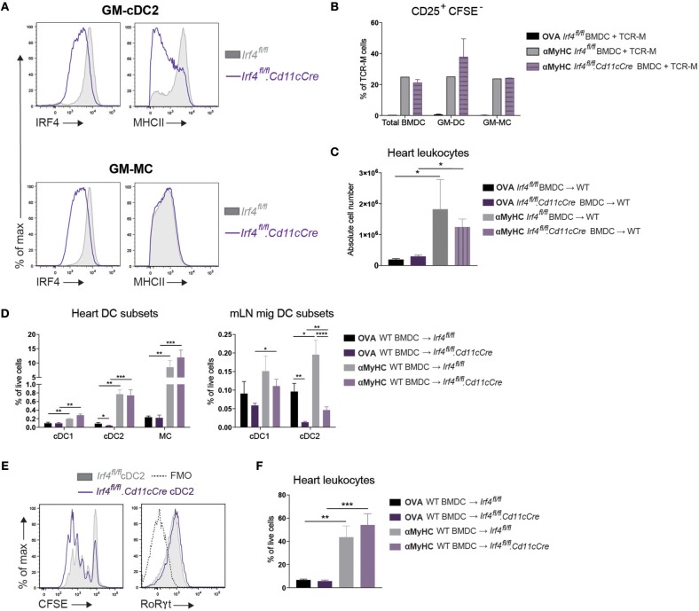
Loss of IRF4 in CD11c+ cells does not affect EAM severity. (A) Histograms representing IRF4 and MHCII expression on GM-cDC2s and GM-MCs from Irf4fl/fl and Irf4fl/fl.Cd11cCre BMDC cultures. (B) CD25+CFSE− TCR-M cell percentages of total TCR-M cells in co-culture with bulk BMDCs, sorted GM-cDC2s, and sorted GM-MCs from Irf4fl/fl and Irf4fl/fl.Cd11cCre BMDC cultures with OVA323−339 peptide as negative control or with αMyHC614−629 peptide. (C) Absolute CD45+ leukocyte number in the heart of WT mice immunized with OVA323−339 or αMyHC614−629 loaded BMDCs cultured from Irf4fl/fl and Irf4fl/fl.Cd11cCre BM. (D) APC subset percentages of total living cells in the heart and mLN of Irf4fl/fl and Irf4fl/fl.Cd11cCre mice immunized with OVA or αMyHC BMDCs (day 10) (E) Histograms of CFSE dilution and RoRγt expression of TCR-M cells co-cultured for 4 days with sorted migratory cDC2s from mLN of Irf4fl/fl and Irf4fl/fl.Cd11cCre mice 10 days after injection with αMyHC BMDCs. (F) CD45+ leukocyte percentage of total living cells in the heart of Irf4fl/fl and Irf4fl/fl.Cd11cCre mice immunized with OVA or αMyHC BMDCs (day 10). All bar graphs in this figure shows data as the mean ± SEM; *P ≤ 0.05; **P ≤ 0.01; ***P ≤ 0.001; ****P ≤ 0.0001.

