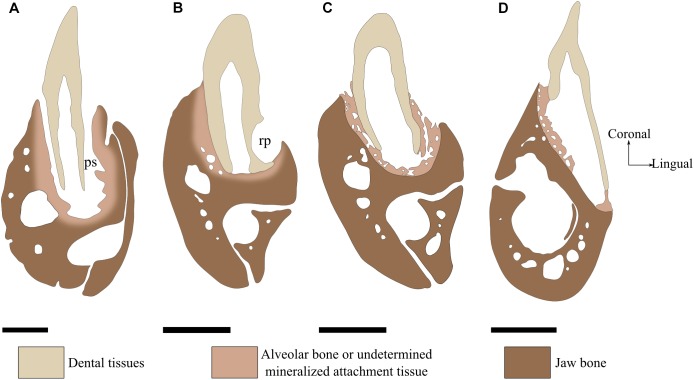
FIGURE 2.
Tooth attachment illustrated by labio-lingual sections of mandibles in various species. (A) Gomphosis attachment associated with a thecodont implantation in Crocodylus niloticus. Note that the tooth and the bone are not in direct contact, leaving a periodontal space (ps). As the limit between alveolar bone and jaw bone is difficult to define, it is drawn as blurred. (B) Ankylosis attachment associated with a subthecodont implantation in Tupinambis teguxin (specimen MNHN 1967-96). The tooth is fused to a mineralized attachment tissue, leaving no periodontal space. Note the presence of a replacement pit (rp). (C) Tooth newly attached in Tupinambis teguxin (same specimen). The “attachment bone” is mineralized, which makes it more clearly distinguishable from the dentary bone. (D) Ankylosis attachment associated with a pleurodont implantation in Cyclura cornuta (specimen MNHN 1919-45). Scale bar is 2 mm.

