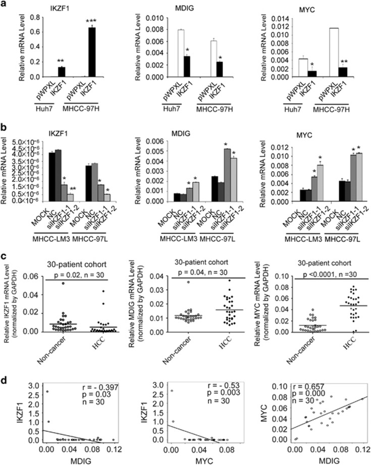
Figure 1.
The expression of IKZF1 was negatively associated to those of MYC and MDIG in HCC. (a) qRT-PCR analysis showed that reduced MDIG and MYC mRNA expression (middle and right panel) in Huh7 and MHCC-97H cells after IKZF1 ectopic expression (left panel). GAPDH was used for normalization control. The graph is shown as mean±s.d. of n=3 independent experiments. Unpaired Student’s t-tests were used for statistical analysis (*P<0.05, **P<0.01, ***P<0.001). (b) Effect of IKZF1 silencing (left panel) on MDIG and MYC mRNA levels (middle and right panel) in MHCC-LM3 and MHCC-97 L cells were determined by qRT-PCR and normalized to GAPDH. The graph is shown as mean±s.d. of n=3 independent experiments. Unpaired Student’s t-tests were used for statistical analysis (*P<0.05, **P<0.01). (c) mRNA levels of IKZF1(left panel), MDIG (middle panel) and MYC (right panel) in a cohort of 30 pairs of human primary HCC patients; HCC, cancer tissue; non-cancer, matched adjacent non-cancerous liver tissues. The data were normalized to GAPDH. Unpaired t-tests were used for statistical analysis (mean±s.d., n=30, *P<0.05, ***P<0.001). (d) The correlation of IKZF1, MDIG and MYC mRNA expression was analysed in 30 primary human HCCs. Pearson’s correlation coefficient (r) and statistical significance are indicated. *P<0.05, **P<0.01, ***P<0.001

