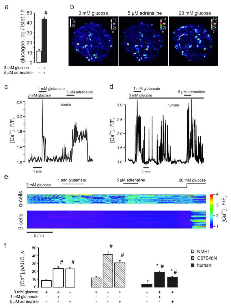
Figure 1. The stimulatory effect of adrenaline on glucagon secretion is mediated by selective elevation of [Ca2+]i in pancreatic α-cells.
(a) Glucagon secreted from isolated NMRI mouse islets in response to 3mM glucose with/without 5μM adrenaline. #p<0.05 vs the effect of 3 mM glucose alone. (b) Variance of the Fluo4 intensity when the islet was perifused with 3mM glucose ± 5μM adrenaline or 20mM glucose (as indicated). The brighter cells are those in which [Ca2+]i oscillates. The arrow indicates a cell that started spiking after adrenaline had been applied. (c-d) Typical single α-cell responses to application of 1mM glutamate and 5μM adrenaline recorded in mouse (c; n=29) and human (d; n=55) islets, at 3mM glucose. (e) Representative [Ca2+]i timecourse in the populations of α- (n=21) and non-α-cells (mostly, β-cells, n=75), differentiated by the response to glutamate. The difference in magnitude of the glutamate and the adrenaline effects was not a consistent finding. (f) [Ca2+]i changes in α-cells quantified as pAUC at 3mM glucose ± glutamate or adrenaline in mouse (NMRI, C57Bl/6N) and human islets. p<0.05 vs the respective effect observed in NMRI mice (*) or the effect of the basal (3 mM glucose) in the same recording (#).

