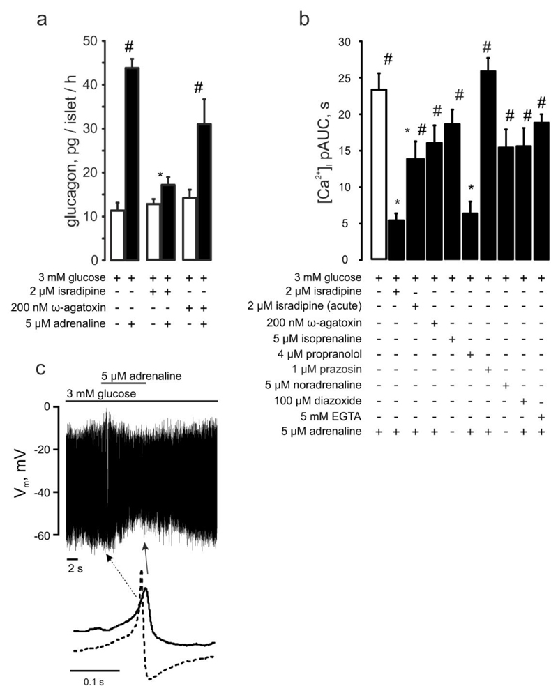
Figure 2. Adrenaline’s effect in pancreatic α-cells depends on Ca2+ influx through L-type Ca2+ channels and is mediated by β-adrenergic signaling.
(a) Glucagon secreted from isolated NMRI mouse islets in response to 3 mM glucose with(out) 5μM adrenaline ± isradipine or ω-agatoxin (as indicated). p<0.05 vs the effect of adrenaline + 3mM glucose (*) or vs the basal (3mM glucose + respective antagonist) group. (b) [Ca2+]i upon adrenergic (isoprenaline (n=27), or noradrenaline (n=19)) stimulation alone (n=29) or with (as indicated) isradipine (n=17, preincubated, n=29, acute), ω-agatoxin (n=13), propranolol (n=11), prazosin (n=49), diazoxide (n=20), EGTA (n=84). p<0.05 vs. the effect of adrenaline alone (*) or vs the basal (3 mM glucose+respective (ant)agonist) of the same recording. (c) Effect of adrenaline on α-cell action potential firing (representative of 11 experiments). Examples of action potentials recorded in the absence and presence of adrenaline (taken from the recording above as indicated) are shown.

