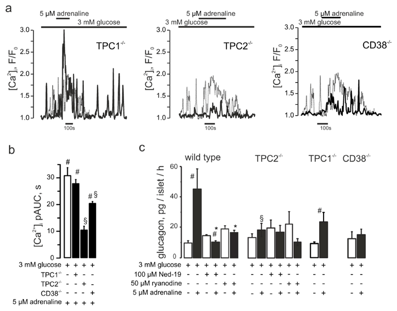
Figure 5. CD38 and TPC2 but not TPC1 mediate the adrenaline response in α-cells.
(a) Effect of adrenaline on [Ca2+]i in α-cells within islets isolated from TPC1-/- or TPC2-/- mice as indicated. (b) [Ca2+]i upon adrenaline stimulation in wild-type, TPC1-/- (n=84), TPC2-/- (n=27) or CD38-/- (n=153) mouse α-cells. p<0.05 vs basal (3 mM glucose) of the same recording (#) or vs the effect of 3 mM glucose + adrenaline (*). (c) Glucagon secretion from isolated mouse islets in response to low glucose (3mM) or adrenaline in the absence/presence of Ned-19 or ryanodine, measured in wild-type C57Bl/6n and TPC1-/-, TPC2-/- and CD38-/- mice. p<0.05 vs the basal (3 mM glucose) (#), vs the effect of 3 mM glucose + adrenaline within the same genotype (*) or vs the effect of 3mM glucose + adrenaline in wild-type animals (§).

