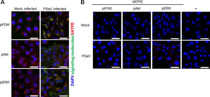
FIG 14.
Direct interaction of pPI3K and pERK with subunit E of the V-ATPase V1 domain as determined by immunofluorescence assay and the Duolink proximity ligation assay. (A) Serum-starved LLC-PK cells were either mock incubated or incubated with the PSaV Cowden strain (MOI = 1 FFU/cell) in the presence of 200 μM GCDCA. Subsequently, the cells were fixed, permeabilized, and incubated with a mixture of primary mouse anti-V-ATPase E subunit and rabbit anti-pPI3K, -pAkt, or -pERK antibody overnight at 4°C. The cells were then incubated with irrelevant secondary antibodies for 1 h at room temperature and processed for confocal microscopy. (B) Serum-starved LLC-PK cells were either mock incubated or incubated with the PSaV Cowden strain (MOI = 1 FFU/cell) in the presence of 200 μM GCDCA. Subsequently, the cells were fixed, permeabilized, and incubated with or without a mixture of primary mouse anti-V-ATPase E subunit and rabbit anti-pPI3K, -pAkt, or -pERK antibody overnight at 4°C. The Duolink PLA was performed as described in Materials and Methods, and the signals are represented by red dots. Representative images are shown. Bars, 20 μm.

