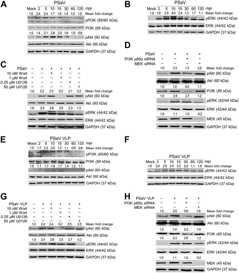
FIG 3.
Activation of PI3K/Akt and MEK/ERK signaling pathways by direct interaction of PSaV in the absence of GCDCA. (A and B) LLC-PK cells were incubated with PSaV (MOI of 1 FFU/cell) in the absence of GCDCA (bile acid) and then harvested at the indicated time points. The levels of PI3K, Akt, ERK, pPI3K p85 (Tyr458)/p55 (Tyr199), pAkt (Ser473), pERK (Thr202/Tyr204), and GAPDH were evaluated by Western blotting using specific antibodies against the target proteins. GAPDH was used as a loading control. (C and D) LLC-PK cells were mock pretreated or pretreated with wortmannin (PI3K inhibitor) or U0126 (MEK inhibitor) at the indicated doses for 1 h at 37°C (C) or transfected with or without siRNAs against PI3K p85α or MEK (D) and then infected with or without PSaV in the absence of GCDCA. Cell lysates were harvested at 5 mpi. The expression levels of pAkt (Ser473), Akt, pERK (Thr202/Tyr204), ERK, and GAPDH were evaluated by Western blotting. GAPDH was used as a loading control. (E and F) LLC-PK cells were incubated with PSaV VLPs (10 μg/ml), and the cell lysates were harvested at 5 mpi and prepared for Western blotting as described above. (G and H) LLC-PK cells were mock pretreated or pretreated with wortmannin or U0126 at the indicated doses for 1 h at 37°C (G) or transfected with or without siRNAs against PI3K p85α or MEK (H) and then incubated with or without PSaV VLPs. Cell lysates were harvested at 5 mpi. The expression levels of pAkt (Ser473), Akt, pERK (Thr202/Tyr204), ERK, and GAPDH were evaluated by Western blotting. GAPDH was used as a loading control. The intensity of each target protein relative to that of GAPDH was determined by densitometric analysis and is indicated above each lane.

