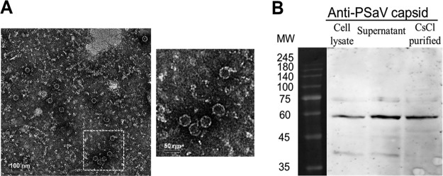
FIG 4.
Electron micrograph and Western blot analyses of PSaV Cowden strain VLPs. (A) PSaV VLPs harvested from recombinant baculovirus-infected Sf9 cells at 72 h postinfection were purified by CsCl density gradient ultracentrifugation and visualized by negative staining with 3% phosphotungstic acid (pH 7.0) under an electron microscope. The inset shows a higher magnification of the left panel. (B) Cell lysates of recombinant baculovirus-infected Sf9 cells at 72 h postinfection, their supernatants, and CsCl-purified VLPs were separated by sodium dodecyl sulfate-polyacrylamide gel electrophoresis, and VLPs were detected by Western blot analysis using anti-PSaV capsid hyperimmune antisera.

