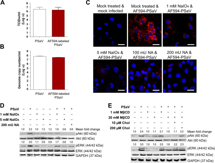
FIG 6.
Activation of PI3K/Akt and MEK/ERK signaling pathways by interaction of PSaV with cell surface carbohydrate receptors. (A and B) LLC-PK cells were infected with mock-labeled PSaV particles or Alexa Fluor 594 (AF594)-labeled PSaV particles and incubated for 36 h at 37°C in the presence of 200 μM GCDCA. The cells were then harvested by freezing and thawing, and the virus titers were determined by TCID50 assay (A) and the genome copy number by real-time RT-PCR (B) as described in Materials and Methods. (C) LLC-PK cells were treated with or without 1 or 5 mM sodium periodate (NaIO4) or 100 or 200 mU Vibrio cholerae neuraminidase (NA) to remove carbohydrates or sialic acids from the cell surface, respectively, incubated with AF594-labeled PSaV particles (approximately 415 particles per cell) for 30 min at 4°C in the absence of 200 μM GCDCA, and subsequently examined by confocal microscopy. This experiment was repeated three independent times, and one representative set of results is shown. Bars, 20 μm. (D and E) LLC-PK cells were treated with or without 1 or 5 mM NaIO4 or 200 mU NA for 1 h at 37°C (D) or treated with 1 or 20 mM MβCD for 1 h at 37°C or with 10 or 200 µM soluble cholesterol for 30 min at 37°C to examine the effect of cholesterol replenishment following MβCD-mediated depletion (E), followed by infection with PSaV (MOI of 1 FFU/cell) in the presence of 200 μM GCDCA. Cell lysates were harvested at 5 mpi. Antibodies against pAkt (Ser473), Akt, pERK (Thr202/Tyr204), ERK, and GAPDH were used to evaluate the expression level of each target protein by Western blotting. GAPDH was used as a loading control. The intensity of each target protein relative to that of GAPDH was determined by densitometric analysis and is indicated above each lane.

