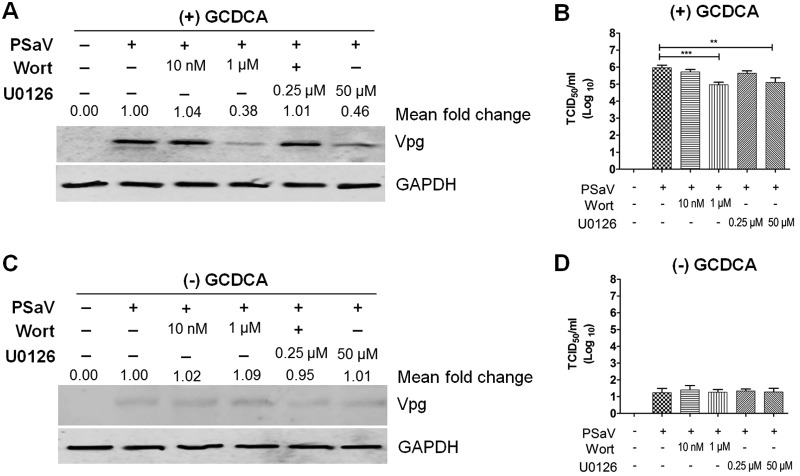
FIG 8.
Inhibition of the PI3K/Akt and MEK/ERK signaling pathways affects PSaV infectivity and viral protein expression. (A to D) LLC-PK cells were pretreated with noncytotoxic concentrations of wortmannin or U0126 for 1 h at 37°C and then infected with PSaV (MOI of 1 FFU/cell) for 36 h in the presence (A and B) or absence (C and D) of 200 μM GCDCA. (A and C) Levels of PSaV VPg protein were determined by Western blotting. GAPDH was used as a loading control. The intensity of VPg relative to that of GAPDH was determined by densitometric analysis and is indicated above each lane. (B and D) Viral titers were determined by TCID50 assay. The data are presented as means and standard deviations of the results of three independent experiments. Differences were evaluated using one-way analysis of variance. *, P < 0.05; **, P < 0.01; ***, P < 0.001; ****, P < 0.00001.

