FIG 9.
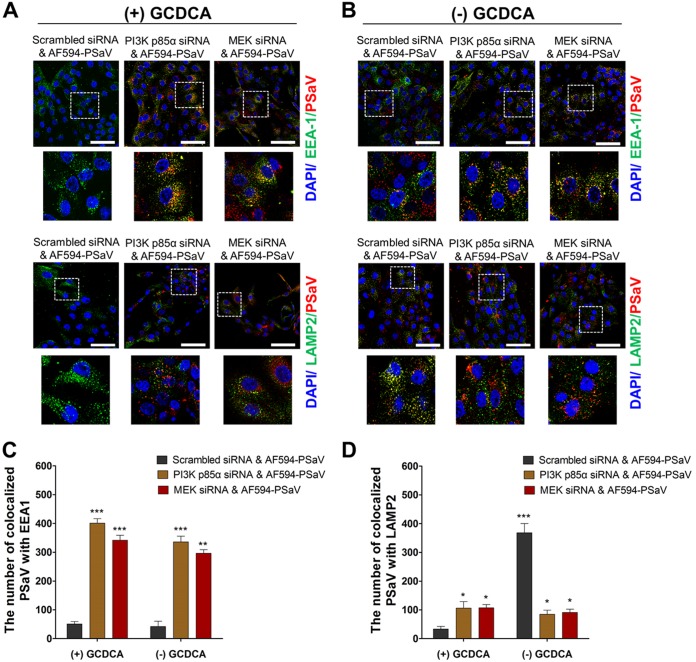
Silencing of PI3K and MEK traps PSaV particles in early endosomes. (A and B) LLC-PK cells were transfected with scrambled siRNA or siRNA against PI3K p85α or MEK and then incubated with Alexa Fluor 594 (AF594)-labeled PSaV particles (approximately 415 particles per cell) for 3 h in the presence (A) or absence (B) of 200 μM GCDCA. After fixation and permeabilization, the cells were incubated with a monoclonal antibody against the early endosome marker EEA1 or the late endosome marker LAMP2 and then with a FITC-conjugated secondary antibody and processed for confocal microscopy to determine the colocalization of AF594-labeled PSaV particles with EEA1 or LAMP2. The boxed areas are magnified and shown under each panel. All experiments were performed in triplicate, and a representative set of results is shown. Bars, 20 μm. (C and D) Quantification of AF595-labeled PSaV particles colocalized with the early endosome marker EEA1 (C) and the late endosome marker LAMP2 (D) was performed using 10 confocal microscopy images of cells treated under the conditions described above by use of the ImageJ program. Quantification of signals was made with a threshold of 0.03 to 1.3 µm2 as described in Materials and Methods.

