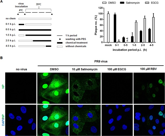
FIG 2.
Effects of salinomycin on the early stages of the influenza virus life cycle. (A) Time-of-addition experiments. The experimental process is described on the left. MDCK cells were infected with influenza PR8 virus for 1 h at 4°C. After removal of unadsorbed virus, the cells were incubated for an additional 4 h at 35°C. They were inoculated under different conditions, i.e., in the absence or presence of 10 µM salinomycin or EGCG. In parallel, at 1, 2, and 4 h p.i., the compounds were added to the cell culture medium. At 5 h p.i., the cell monolayers were washed with PBS and overlay medium was added to allow plaque generation. The numbers are expressed as percentages relative to plaque number from the DMSO-treated sample and represent the means ± SD of triplicate samples. (B) Confocal microscopy showing the subcellular distribution of viral NP. MDCK cells were mock infected (no virus) or infected with PR8 virus at an MOI of 2.5 for 4 h at 37°C in the presence of DMSO, salinomycin, EGCG, or RBV. The viral NP protein was detected using an NP-specific monoclonal antibody and an Alexa Fluor 488-conjugated goat anti-mouse secondary antibody (green). Nuclei were counterstained with DAPI (blue). Original magnification, ×400.

