
FIG 6.
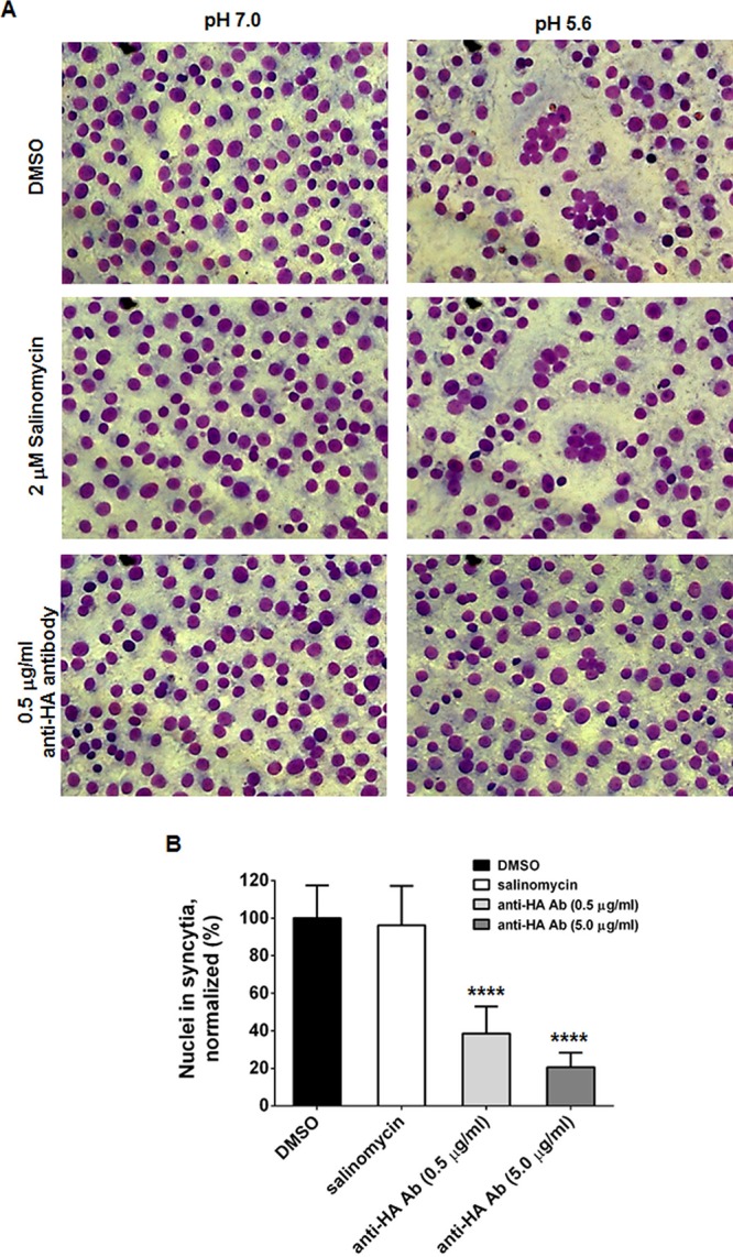
Membrane fusion of cells infected with PR8. (A) Vero E6 cells were infected with PR8 at an MOI of 0.5 at 37°C. At 16 h p.i., cells were preincubated with TPCK-trypsin (5 μg/ml) together with DMSO, 2 μM salinomycin, or 0.5 μg/ml anti-HA2 antibody. Cellular membrane fusion was initiated by exposing samples to the indicated conditions (pH 7.0 or 5.6). After staining with Giemsa, fixed cells were visualized by microscopy. Original magnification, ×200. (B) The relative number of nuclei in syncytia was counted from 16 representative images per sample at pH 5.6. Statistical significance was analyzed by comparing differences between the DMSO-treated group and the compound- or antibody-treated groups. ****, P < 0.0001.