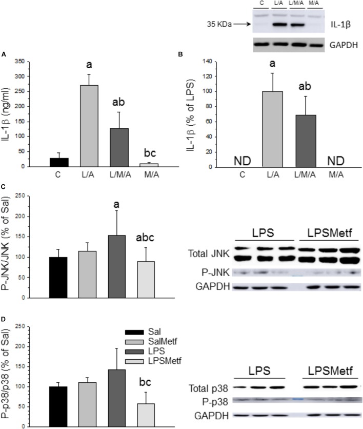
FIGURE 6.
Effect of metformin and LPS on inflammasome activation in BV2 cells and expression of JNK, P-JNK, p38, and P-p38 proteins in the SN. (A) Extracellular IL-1β measured by ELISA. (B) Intracellular IL-1β measured by Western blot. Results are mean ± SD of three independent experiments for all groups, expressed as ng/ml (A), and three independent experiments for all groups, expressed as percentage of the L/A group (B); intracellular amounts are normalized to GAPDH expression. Statistical significance (one-way ANOVA followed by the LSD post hoc test for multiple comparisons): a, compared with the control group; b, compared with the L/A group; c, compared with the L/M/A group; p < 0.001. (C) Control group; L/A, cells treated with LPS and ATP; L/M/A, cells treated with a combination of LPS, metformin and ATP; M/A, cells treated with a combination of metformin and ATP; ND, not detected. Proteins from the SN of rats under the different treatments assayed were separated by electrophoresis and transferred to nitrocellulose membranes, and stained using anti-JNK, anti-P-JNK, anti-p38 and anti-P-p38 antibodies. Total optical density of each band was calculated. Results are mean ± SD of seven independent experiments for groups Sal, LPS and LPSMetf and eight independent experiments for group SalMetf (C), and three independent experiments for groups Sal and LPS and four independent experiments for groups SalMetf and LPSMetf (D), expressed as P-JNK/JNK (C) and P-p38/p38 (D) intensity ratios (normalized to GAPDH expression) relative to the control (Sal) group. Statistical significance (one-way ANOVA followed by the LSD post hoc test for multiple comparisons): a, compared with the control (Sal) group; b, compared with the SalMetf group; c, compared with the LPS group; p < 0.01.

