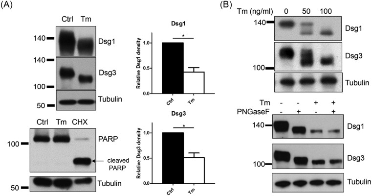
Figure 3. Dsg1 and 3 are reduced in amount and molecular weight, showing non-glycosylated proteins in NHEKs by treatment with tunicamycin.
(A) Confluent NHEKs were pretreated with 100 ng/ml tunicamycin for 8 h before incubating the cells in a high calcium medium (1.3 mM) with tunicamycin for an additional 16 h. Treatment with cycloheximide (100 μg/ml) for 24 h was used as a positive control for apoptosis. Western blot analyses were performed for Dsg1 and 3. Data were depicted as means ± S.D., n=4; *P<0.05. (B) Incomplete N-glycosylation inhibition was examined using different doses of tunicamycin (50 or 100 ng/ml). Deglycosylation was confirmed with PNGaseF. PNGase F reaction was performed through Bio-Rad kit according to the manufacturer’s instructions. Cell lysates were prepared with RIPA buffer. Abbreviations: CHX, cycloheximide; Ctrl, Control; Tm, tunicamycin.

