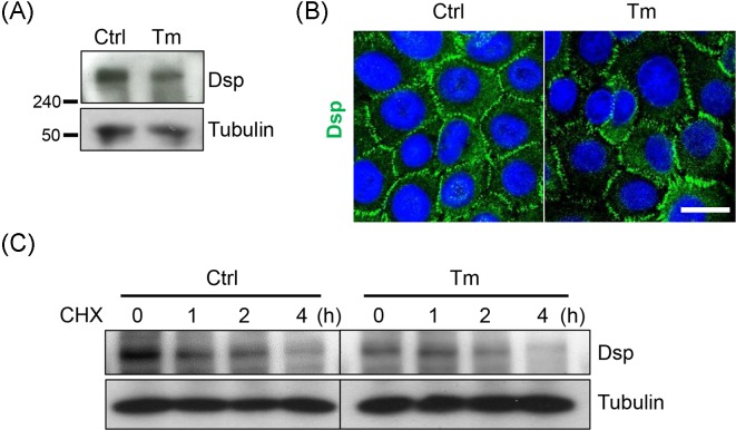
Figure 8. Dsp appears normal distribution at the cell borders and comparable stability despite tunicamycin treatement.
Confluent NHEKs were pretreated with or without 100 ng/ml tunicamycin for 8 h then switched to a high calcium medium (1.3 mM) for an additional 16 h. (A) Dsp was detected through Western blotting. (B) Cells were prepared on the coverslips. Immunostaining was performed to detect Dsp. Scale bar = 10 μm. (C) After treatment of tunicamycin and calcium, cycloheximide (30 μg/ml) was added, and the cells were harvested at 0, 1, 2, and 4 h after the cycloheximide treatment. Abbreviations: CHX, cycloheximide; Ctrl, control; Tm, tunicamycin.

