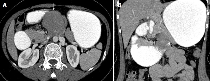
Figure 1.

Oral and intravenous contrast-enhanced multidetector computed tomography. A: Axial cross-sectional views of the multidetector computed tomography (MDCT) scan; B: Coronal reformant cross-sectional views of the MDCT scan. A space occupying mass lesion with homogenous density showing minimal contrast uptake is seen in the preaortic area in the abdominal midline (white arrow). Coronal reformant MDCT images show that the mass is in the fourth part of the duodenum (curved black arrow). There is only slight oral contrast passage to jejunal loops (thin black arrow), and the duodenum and stomach had a ptotic appearance due to mechanical obstruction caused by the mass.
