Figure 6.

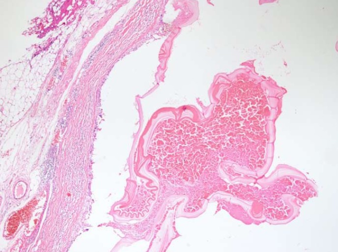
Microscopic appearance of the hydatid cyst tissue stained with hematoxylin and eosin. An acellular membrane of a hydatid cyst is shown here (HE × 40).

Microscopic appearance of the hydatid cyst tissue stained with hematoxylin and eosin. An acellular membrane of a hydatid cyst is shown here (HE × 40).