Figure 5.
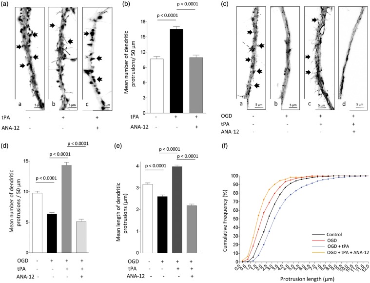
Treatment with rtPA protects the post-synaptic terminal via TrkB receptor activation. (a) and (b) Representative micrographs (a) and mean number of dendritic protrusions/50 µm (b) from Wt neurons incubated during 5 min with either PBS (a), or 5 nM of tPA, alone (b) or in combination with 30 μM of ANA-12 (c). n = 30 neurons per condition from three different cultures. Lines denote SEM. Arrows in a – c denote dendritic spines and filopodia. (c) and (d) Representative micrographs (c) and mean number of dendritic protrusions/50 µm (d) in Wt neurons maintained under normoxic conditions (a), or exposed to oxygen and glucose deprivation (OGD) conditions, alone (b) or in the presence of 5 nM of tPA and either PBS (c), or ANA-12 (d). Arrows in (c) (panels a and c) denote dendritic filopodia. n = 50 neurons per condition from three different cultures. (e) and (f) Mean length of dendritic protrusions (e) and cumulative frequency of spine length (f) in Wt neurons exposed to OGD conditions and treatment with rtPA and ANA-12 as described in (c) and (d). n = 120 neurons per experimental condition.

