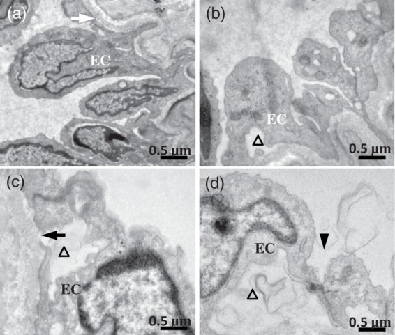
Figure 3.

The effect of maternal HFD consumption on ultrastructure appearance of the MCAs. (a) C/C group. The normal base membrane (white arrow) and endothelial cell (EC) were observed. (b) Showing the endothelial damage and endothelial denudation (triangle) in HFD/C rats. (c) C/HFD group. Endothelial cellular swelling, disintegrated basal lamina (dark arrow), and endothelial denudation (triangle) were observed. (d) Showing obvious endothelial denudation, swollen endothelial nucleus, and enlarged intercellular junction (dark arrowhead) in HFD/HFD offspring. n = 3 for each group. Scale Bar: 0.5 µm.
