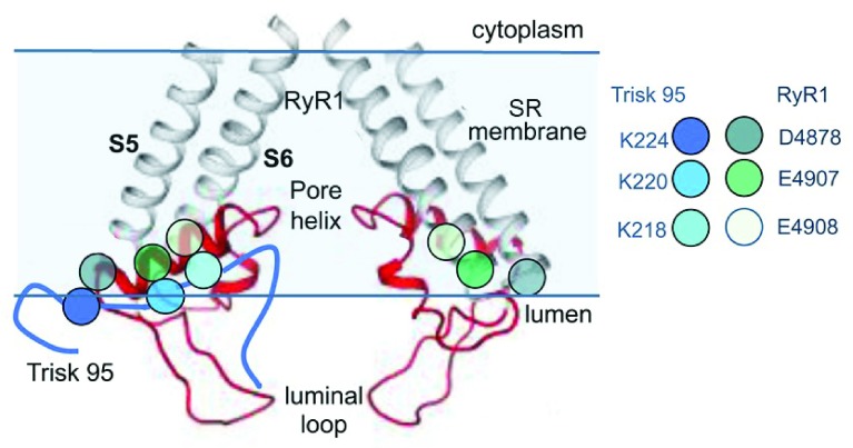
Figure 3. Proposed region for ionic interactions between skeletal triadin (Trisk 95) and RyR1.
The near-atomic resolution structure of the pore-forming elements of RyR1 showing two diagonal protomers (Extended Data Figure 8 in 10). The grey transmembrane S5 and S6 helices and red pore helix, S5–pore helix linker, and SF linker between the pore helix and S6 helix are shown. Mutagenesis studies suggest that residues D4878, E4907, and E4908 in the outer regions of the pore helix are associated with K218, K220, and K224 in Trisk 95 39, 40. The approximate positions of E4907 and E4908 are indicated by the black arrowhead, and the arrow indicates the predicted binding site for Trisk 95. RyR, ryanodine receptor; SR, sarcoplasmic reticulum. Reprinted by permission from Springer Nature: Pflügers Archiv European Journal of Physiology, Three residues in the luminal domain of triadin impact on Trisk 95 activation of skeletal muscle ryanodine receptors, E. Wium, A. F. Dulhunty, N. A. Beard, © 2016.

