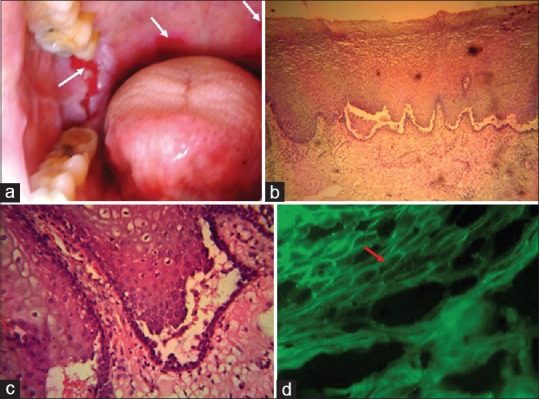
Figure 3.

Multiple painful ulcers in right buccal mucosa and soft palate (a). Stained Haematoxylin and Eosin sections [×400] revealed stratified squamous epithelium with intraepithelial blister formation, characteristic suprabasilar cleavage, and presence of acantholytic large, rounded keratinocytes with a hypertrophic nucleus and a perinuclear halo - “Tzanck cells” (c) in the split area. DIF revealed intercellular substance deposition of IgG, : characteristic “fish-net appearance” (d).
