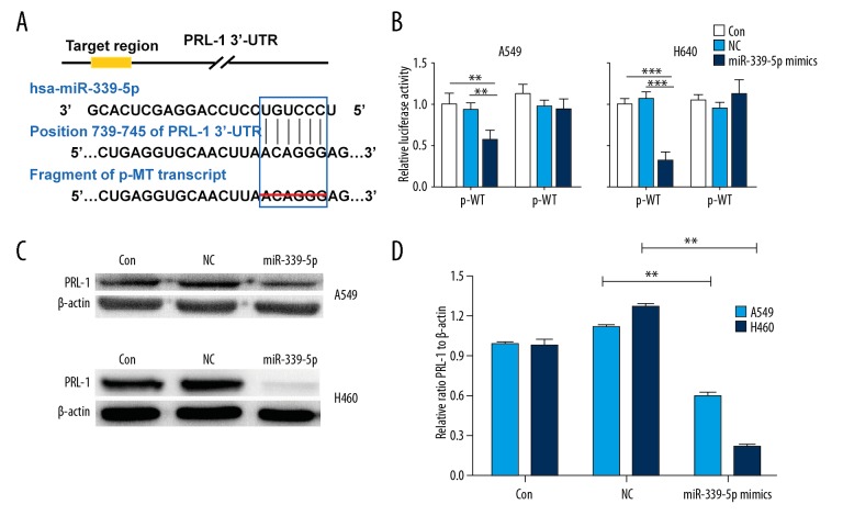
Figure 5.
PRL-1 is a target of miR-339-5p in lung cancer cells. (A) Predicted miR-339-5p binding sites in the 3′-UTR of PRL-1 by TargetScan and the designed mutant 3′-UTR in which miR-339-5p binding sites were deleted. (B) Luciferase reporter assays in A549 and H460 cells transfected with p-WT or p-MT alone (Mock) and cotransfected with p-WT or p-MT and NC or miR-339-5p mimics; p-WT=the reporter plasmid of wild-type PRL-1 3′-UTR, p-MT=the reporter plasmid of mutant-type PRL-1 3′-UTR. Data are normalized according to the ratio of Firefly luciferase activity to Renilla luciferase activity. (C) The expression levels of PRL-1 were detected by Western blotting in A549 and H460 cells after indicated transfections. β-actin was used as a loading control. (D) Quantification of Western blotting results as in (A). Data are expressed as the mean ±SD (** P<0.01, *** P<0.001).

