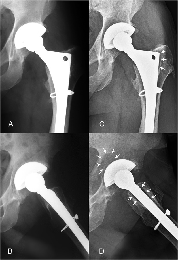
Fig. 3.

The patient with XLPE who had osteolysis exceeding 1.5 cm2 in the current study demonstrated no evidence of osteolysis at 8-year followup on AP (A) or lateral (B) radiographs. At 16-year followup, proximal femoral osteolysis (designated by the white arrows) was noted on the AP (C) and lateral radiographs (D). An acetabular lesion behind the dome hole with an area of 2.9 cm2 was also noted on the lateral view (D). The patient was a man who was 43 years of age at the time of his primary THA and who had a head penetration rate of 0.07 mm/year and a linear wear rate of 0.05 mm/year.
