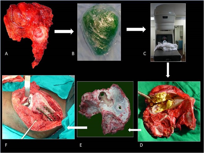
Fig. 1 A-F.
The ECRT technique. (A) The specimen is resected as per oncologic principles after which (B) the specimen is wrapped in vancomycin-saline mops and plastic wrap to maintain sterility while being transported to the radiation department. (C) Radiotherapy: single-fraction 60 Gy with 6-mV photons at 3 Gy/min is delivered to the specimen. (D) The specimen is received on a separate sterile trolley, stripped of all soft tissue, and (E) cleaned thoroughly with vancomycin mixed with saline. (F) The specimen is fixed with 3.5-mm reconstruction plates. Resurfacing with a cemented constrained liner is done for tumor invasion into acetabulum.

