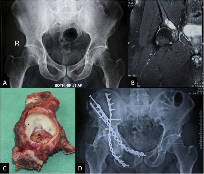
Fig. 3 A-D.
A 43-year-old man with periacetabular chondrosarcoma treated with ECRT. The patient presented with hip pain. (A) Preoperative radiograph appears normal and the lesion is not visible, whereas (B) MRI shows the periacetabular lesion, which was diagnosed to be a Grade 2 chondrosarcoma by CT-guided biopsy. (C) Resected specimen was treated with ECRT and reimplantation with transiliac screws and two-column fixation as seen on the (D) 4-year postoperative radiograph showing the healed junctions. There is heterotopic bone, which did not cause any restriction of movement. The joint space is good. This man walks without a limp and is back to working full-time.

