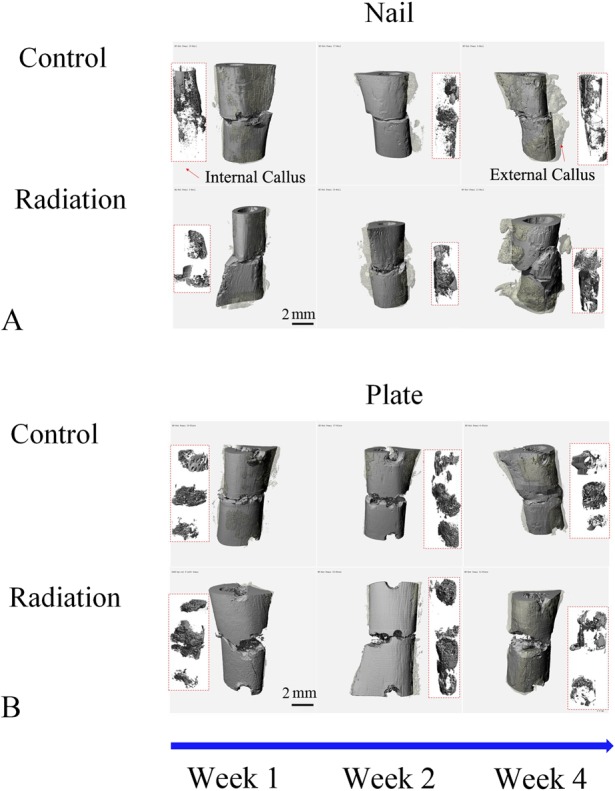
Fig. 2 A-B.

These images represent 3-D micro-CT reconstructions of the external and internal callus present around the fracture sites in rat femora using either nail (A) or rigid plate (B) fixation at Weeks 1, 2, and 4 postoperatively. The original femur and surrounding external callus are colored with solid and transparent gray, respectively. The internal callus is shown in the rectangle box inset (red outline).
