
Fig. 3 A-D.
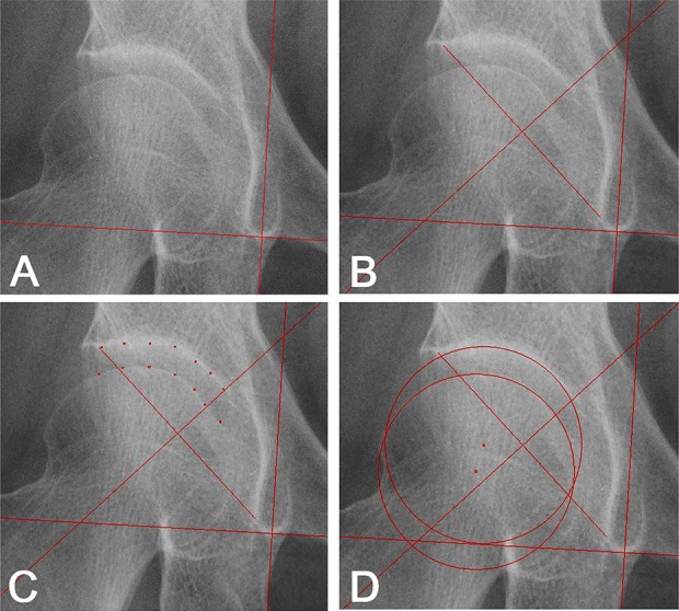
The center gap measurement method is shown. (A) A vertical line is drawn based on the horizontal line connecting the inferior ends of the left- and right-sided teardrops. (B) A perpendicular bisector of a line segment is drawn connecting the inferior and superior edges of the acetabulum. (C) Seven points on the joint surface except the fossa in the superior half of the acetabulum that is separated by the perpendicular bisector are plotted. The surface of the femoral head is plotted using seven points within the same region as the acetabulum. (D) Circles of the acetabulum and femoral head are automatically constructed by the interpolation of those seven points. Distances in the horizontal and vertical planes and the absolute value between the centers are measured digitally. All measurements are normalized using the known diameter of the measure on radiographs.