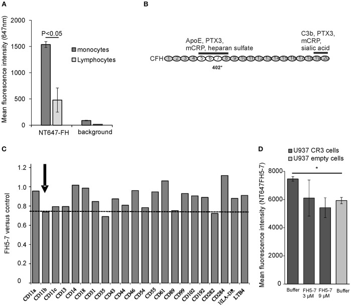
Figure 1.
Factor H binding to peripheral blood leukocytes. (A) Peripheral blood mononuclear cells were incubated with NT647-labeled FH and the mean fluorescence intensities were analyzed by flow cytometry (n = 3). (B) Schematic structure of the full length FH and the location of domains 5–7 that interact with apoE. The binding sites of different ligands, apoE, pentraxin3 (PTX3), monomeric C-reactive protein (mCRP) and heparin sulfate, and the common polymorphic Y402H site on domain 7 are shown. (C) Screening of FH5-7 binding by a panel of antibodies directed against various receptors and surface molecules on monocytes. Peripheral blood monocytic cells were preincubated for 30 min with or without 12 μg/ml of FH5-7 prior adding 2 × 10⁵ cells cells/well in a 96-well plate with FITC-, APC- and/or PE-labeled antibody. Mean fluorescence intensities were calculated from the gated cells using flow cytometry. Inhibition of antibody binding was calculated by dividing the mean fluorescence of FH5-7 treated cells by PBS treated cells (FH5-7 vs. control). The CD11b part of CR3 dimer is marked. The dashed line shows the level of anti-CD11b binding in the presence of FH5-7 (< 0.8) compared to binding of the antibody without FH5-7 (1.0). (D) CR3 expressing and empty U937 mononuclear cells were incubated with NT647 labeled FH5-7 in the presence or absence of unlabeled FH5-7 (x-axis). Statistical significance between multiple samples was calculated using one-way ANOVA supplemented with non-parametric Tamhane's post-hoc multiple comparison test. Error bars indicate SD. * = p < 0.05.

