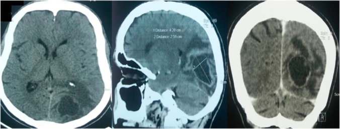
Fig. 1.

Brain CT scan showing intra axial parieto-occipital lesion mesearing 45 mm of long axis which is hypodense and unenhanced in its center with thick enhanced wall and having intensely enhanced left anterolateral focal thickening. This lesion is surrounded by important edema with mass effect on cortical grooves and the left occipiatal horne of the homolateral ventricule.
