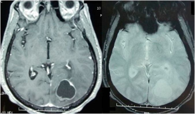
Fig. 2.

Brain MRI showing the occipital inta axial lesion with fluid center hypo intense on T1 weighted image, which presents at the periphery a tissue component of variable thickness that intensely enhances. This lesion comes into contact with the superior sagittal sinus without thrombosis.
