
Fig. 1.
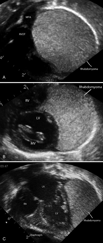
( A ) Short axis subcostal view and right ventricular outflow tract. Rhabdomyoma extending anteriorly around the right ventricle. ( B ) Subcostal short axis view at the level of the mitral valve. Rhabdomyoma surrounding the left ventricle. ( C ) Subcostal apical four chambers. Rhabdomyoma arising from the posterior wall of the left ventricle. Multiple small rhabdomyomas in the left ventricle and mitral valve. LA, left atrium; LV, left ventricle; RA, right atrium; RVOT, right ventricular outflow tract.