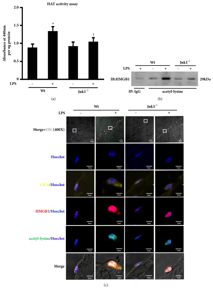
Figure 4.
The role of JNK signal activation in LPS-induced HMGB1 acetylation. (a) Primary peritoneal mesothelial cells from wild-type (Wt) and Jnk1-/- mice were treated with LPS for 48 h, and then examined HAT activity. (b) Cells were treated and described as above. Acetyl-HMGB1 level was examined by immunoprecipitation. (c) Parietal peritoneum in each group was stained for CK18 (yellow), HMGB1 (red), and acetyl-lysine (green). Hoechst (blue) was used for nuclear staining. Scale bar: 5um. Inserts showed particular area at higher magnification to better visualize the location of HMGB1 and acetyl-lysine.

